
Main Index
In Store
Our Web Store
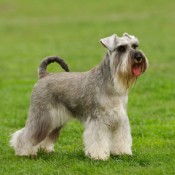
Miniature Schnauzer Picture Gallery
Latest Dog Blogs
- What Are The Basic Commands To Train A Dog?
- PaySafe As The Most Popular Type Of Deposit
- Everything You Need To Know About Pet Sales
- Dogs Contribute To Our Physical And Mental Well Being
- How To Choose Where To Bet On Greyhounds In 2022
- Volunteer With Animals - How To Help Dogs Around The World
- Basic Understanding Of The House Edge
- Why You Should Get A Dog
- Top 20 Popular Dog Names Around The World
- Constipation in Dogs and How to Find Solutions

The Complete Review Of The Structure And Function Of The Skeleton In Dogs
- 01/02/2014
he skeleton of the dog is referred to in two parts:the axial skeleton and the appendic-skeleton. The axial skeleton includes the skull, which varies considerably from breed to breed while retaining the same essential characteristics. The spinal column is also part of the axial structure, and starts with the seven cervical vertebra which form the neck. At the head end, the first vertebra, known as the Atlas, permits the head to move up and down, while the second vertebra, the Axis, enables the head and the Atlas to rotate. The remaining cervical vertebrae allow the dog sufficient flexibility to turn the neck to look directly behind, without moving the body.
Below the neck, thirteen thoracic vertebrae protect the spinal cord and support the thirteen pairs of ribs which form a flexible ribcage to protect the heart and lungs. From the end of the ribcage, seven lumbar vertebrae support the abdomen and lead to the sacrum, which usually consists of three fused vertebrae, followed by a series of caudal vertebrae which form the tail. However, this feature varies greatly from breed to breed.
The appendicular skeleton consists of the forelimbs and hindlimbs. The forelimb has a strong flattened shoulder- blade or scapula, attached by strong muscles to the skeleton. At its lower end it is connected via a highly flexible joint to the humerus-bone. The lower end of humerus-bone is attached to paired bones, the radius and the ulna. Moving further down, the radius and ulna meet the carpal joint. This is equivalent to the human wrist and is made up of several small bones, arranged in two rows, and forming a joint with the foot.
The carpal joint moves mainly by flexion and extension, but is also capable of some rotation. The dog's foot has a series of five metacarpal bones, side by side, and each metacarpal has a corresponding digit consisting of three phalanges, the third of which is covered by a claw. The inside digit is the smallest, and may be absent in some dogs. When present it is known as the dew-claw. The hind limb is attached to the pelvic girdle, which is formed of three paired bones fused into a ring-shaped structure, and attached to the axial skeleton at the sacrum. On either side of the girdle is a depression called the acetabulum which provides a socket for the head of the femur or thigh bone. The lower of the femur articulates with the partly fused tibia and fibula. This joint, known as the stifle joint, is protected by the patella or kneecap. At the lower end of the tibia the hock joint, or tarsal, is equivalent to the carpus in the forelimb, and the hind foot has the same basic structure as the forefoot, though the dew-claw is rarer in the hind-limbs.
Please Help Us
IrishDogs.ie takes a lot of time, money and hard work to produce. But we do it because we believe our perspective matters because it might well be your perspective, too.
Our future could be much more secure with your help. Please SUPPORT us by clicking on the Donate Button at the Top Right of your screen.
Quick Search
Donate
Latest Dog Pods
- Tips on How to Stop Your Dog from Biting
- Beware - Not All Advertised Dog Rescues Really Are! How Can You Know The Truth?
- Helpful Tips For Dog Obedience Problems
- How to Keep Dogs From Eating Poop
- Dog Grooming Tips - A General Overview of the Very Basics of Dog Grooming
- Recognising Different Types of Dog Obedience Problems
- 5 Important Tips On Feeding A Puppy